-and-
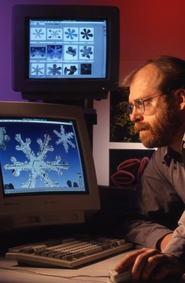
In 1993, the Agricultural Research Service began photographing snowflakes with scanning electron microscopes (SEMs).
Source: National Agricultural Library
Photo: Courtesy
In 1993, the Agricultural Research Service began photographing snowflakes with scanning electron microscopes (SEMs). Microscopists William Wergin and Eric Erbe collected fresh snow from their cars, froze it with liquid nitrogen at -320°F, and covered it with a thin layer of platinum. A SEM passed electrons over the snow and rendered the flakes as images.
SEMs are stronger than light microscopes and can magnify snow crystals 10,000 to 20,000 times their size. Enhanced images allow scientists to better examine snowflakes’ size and structure—both important factors in determining the amount of water in a mountain snowpack. Melting snowpacks replenish reservoirs for public and agricultural use, so information from snowflake micrographs can help scientists predict the summer’s water supply.
Discover more uses for electron microscopy technology in Agricultural Research (now named Tellus), and browse other editions of the USDA’s science magazine in NAL’s digitized collections.


